MICAIA® studio

Smart and rapid Image Analysis for quantitative Results in Microscopy

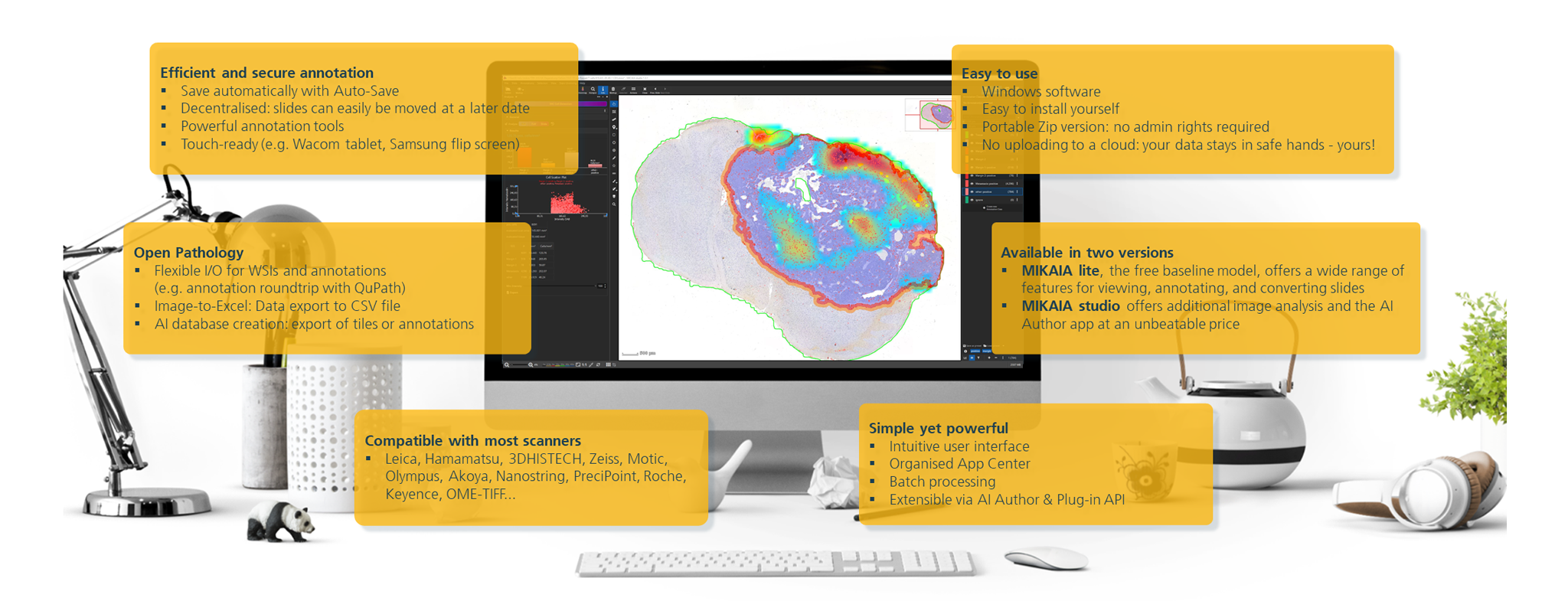
  • Whether as MICAIA® lite, the basic version available free of charge, or as MICAIA® studio, for which there is a range of custom pricing models, MICAIA® is a cost-effective alternative to other software tools.
  • Results are visualized in real time and can be exported as a CSV file. This means they can be evaluated at a glance and used for further data analysis.
  • For each task, whether that involves traditional image processing or deep learning, we select the tool that will yield optimum results.
  • We collaborate with clinical partners to ensure that our software meets each user’s requirements. We view the initial sample images together with them and have them brief us on the biological background and research questions. We translate these questions into an IT problem and design an initial algorithm to solve it. Together, we refine the process over multiple iterations.
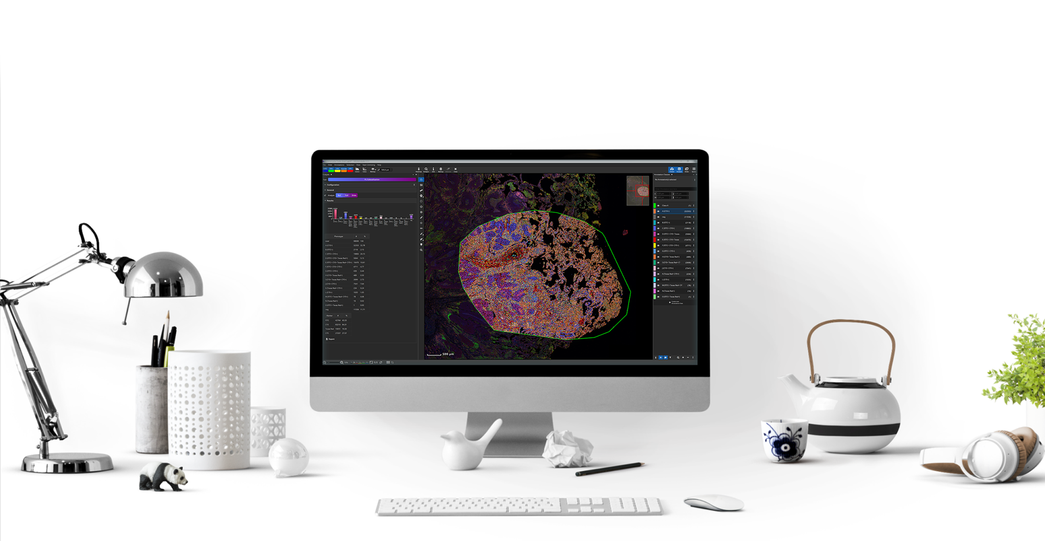
  • MICAIA® studio supports all the usual WSI formats and offers apps for bright-field and fluorescence microscopy.

Straight from research to application: MICAIA® is our platform for transferring the results of our research projects to the sphere of application.

Quantitative Analysis of High-Plex Immunofluorescent Whole-Slides:
White Paper

-----

MICAIA® is distributed by:

  • Smart In Media – The Pathologist's Company
  • Benestar Tech

MICAIA® Studio Preview

AI powered tissue cartography for colorectal cancer. The first half of the video shows the standard version of this App, which divides the slide or ROI into non-overlapping patches and classifies them into 7 tissue classes (tumor cells, healthy mucosa, connective tissue/fat, muscle, necrosis, mucus, inflammation). Each class is assigned a false color. Optionally, a single class can be visualized as a heatmap. The AI’s confidence is mapped into the opacity, respectively heatmap temperature. The second half shows the Fast Tissue Cartography, which uses the same AI model, but first divides the image into small clusters of similar content. Then, a random subset of patches per cluster is analyzed and the entire cluster is assigned to a tissue class using majority voting. After the classification, the tumor outline and invasive margin are derived.

Digital Pathology Projects


Immunoprofiling

DigImmune – coexpression analysis in brightfield using serial sections


Single Cell Detection

Single cell tumor detection in lymph nodes


AI in Digital Pathology

Robustness and explainability


Tumor Budding

Automatic scoring of tumor buds

MICAIA® | YouTube Playlist