PathoScan – Quality Control on Overview Image

PathoScan
© Fraunhofer IIS
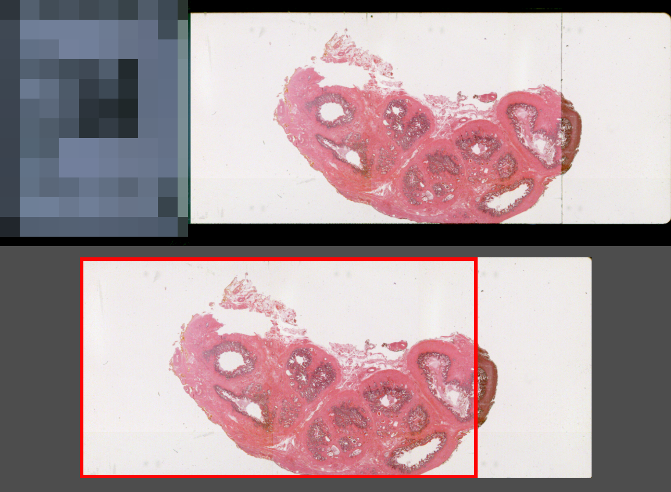
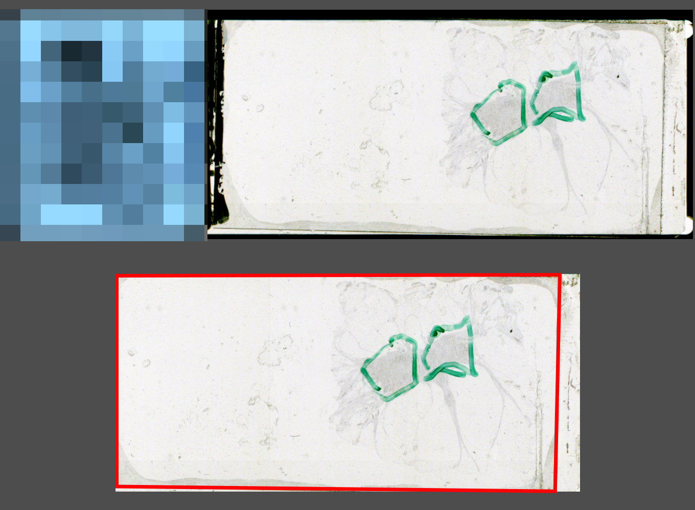
Cover glass does not completely cover the tissue
PathoScan
© Fraunhofer IIS
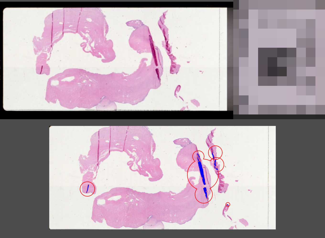
Folds are recognized
PathoScan
© Fraunhofer IIS
Folds are recognized
PathoScan
© Fraunhofer IIS
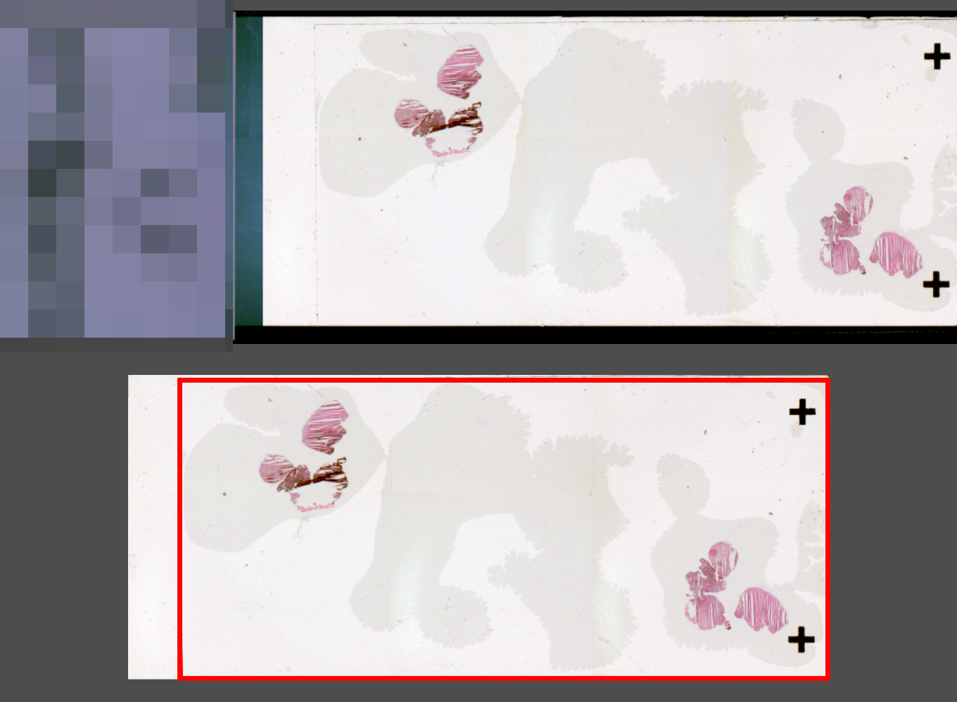
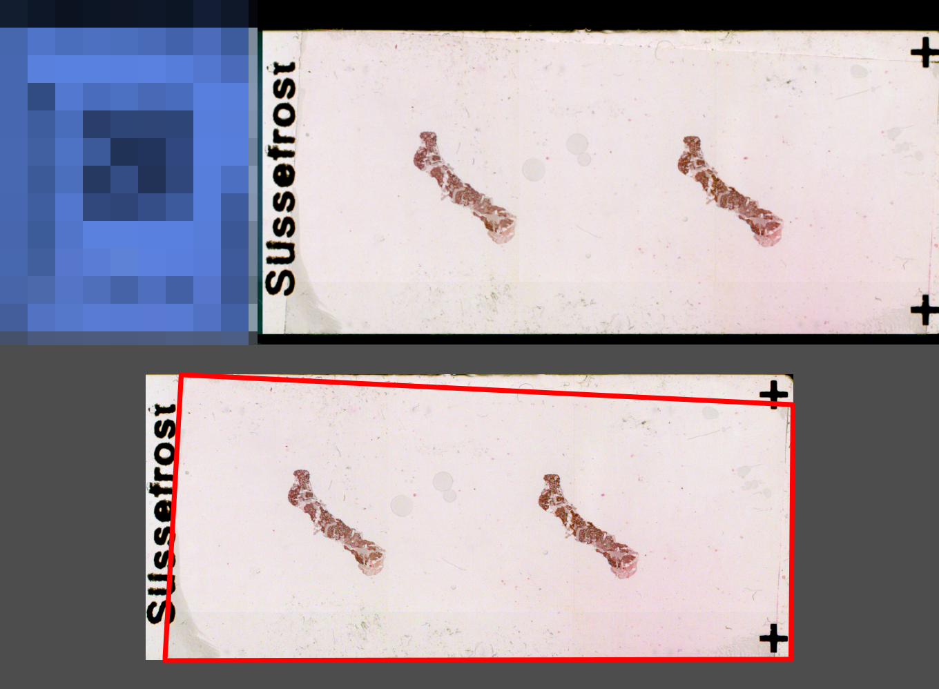
Overlapping cover glass - risk of jamming or glass breakage in the tissue scanner
PathoScan
© Fraunhofer IIS
Overlapping cover glass - risk of jamming or glass breakage in the tissue scanner
PathoScan
© Fraunhofer IIS
Overlapping cover glass - risk of jamming or glass breakage in the tissue scanner

PathoScan project: Automated digitization in routine pathology

Goal: Digital and automated microscopy workflow including pre-scan quality control using an overview camera

Partner: PreciPoint GmbH, University of Regensburg, Technical University of Munich TUM, HTI bio-X GmbH


Current Status:

  • Systematic collection and prioritization of possible error artifacts that can occur during sample preparation
  • Collection and digitization of problematic slides

Database: Slides are currently being collected in Regensburg and Munich and then digitized. Using the database, the scientists at Fraunhofer IIS will develop and validate the image analysis algorithms.

Our Solution:

  • Coloring (HTI Smart Automation) and digitization (PreciPoint) using a fully automated digitization workflow
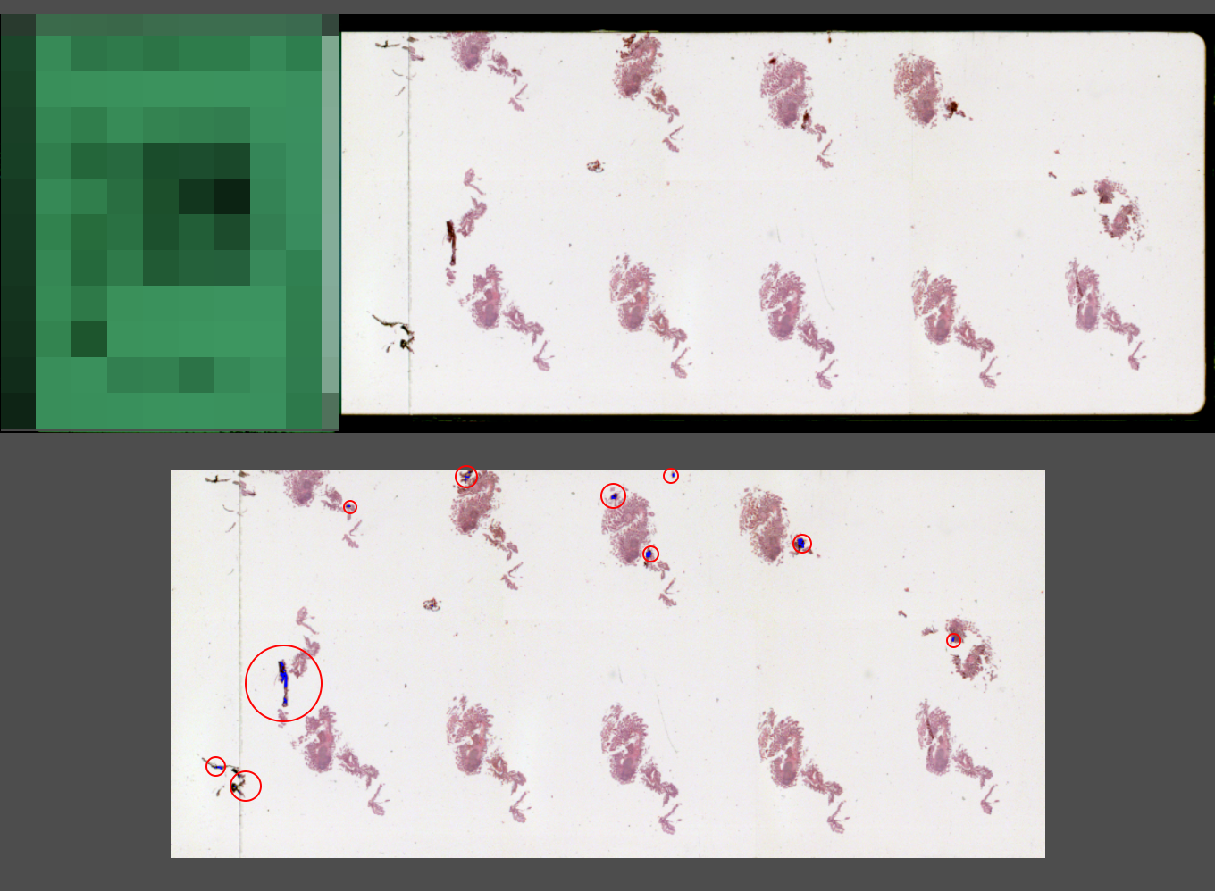
  • Automatic image analysis using the overview camera to detect various quality problems such as protruding cover glasses, air bubbles, foreign particles, folds, cracks
  • Detection of over / under-staining or faulty staining by objective quantification of the coloring
  • Cost and time efficient quality assurance procedures
-------------------------------------------------
Press
 

Tissue Pathology: PathoScan project - Automated digitalization in routine pathology

PreciPoint: Development Project PathoScan: Automated Digitization in Pathology – Joining Forces for Digitization


More Information

Your Development Partner for Medical Image Analysis

Talk to us – together we will find a solution for your medical image processing challenge.

Software based Image Analysis and Enhancement

Our focus are the domains of Microscopy and Endoscopy.

Medical Image Analysis – Publications

Here you will find our publications arranged by year.