Fraunhofer Institute for Integrated Circuits IIS
See.Feel.Train.
HandsOn.surgery is a surgical simulator that addresses all senses. The training is carried out with individualized 3D models from patients segmented from real volumetric image data (e.g. CT, DVT, MRI).
HandsOn.surgery is a surgical simulator that addresses all senses. The training is carried out with individualized 3D models from patients segmented from real volumetric image data (e.g. CT, DVT, MRI).
See |
|
Hear |
|
Feel |
|
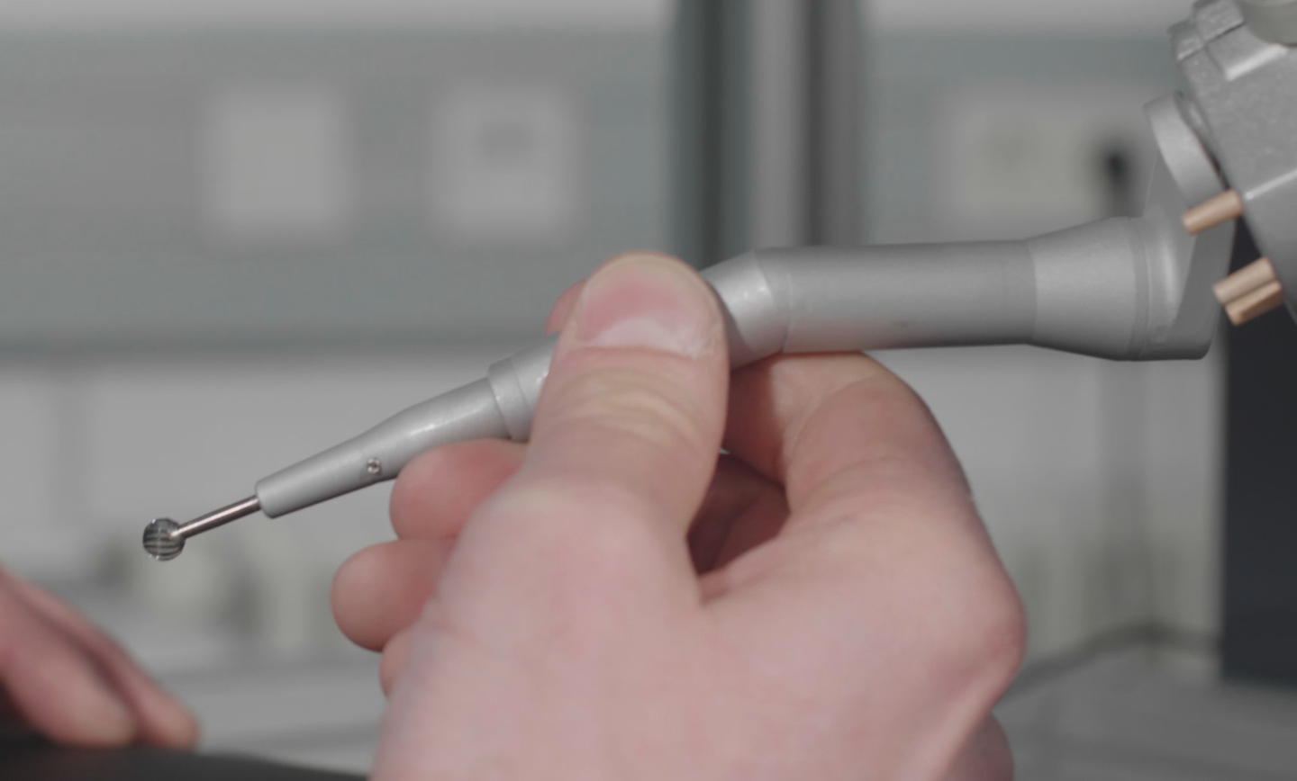
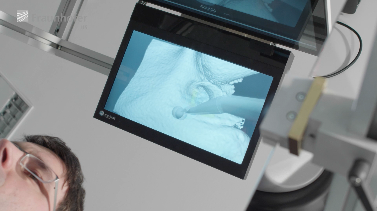
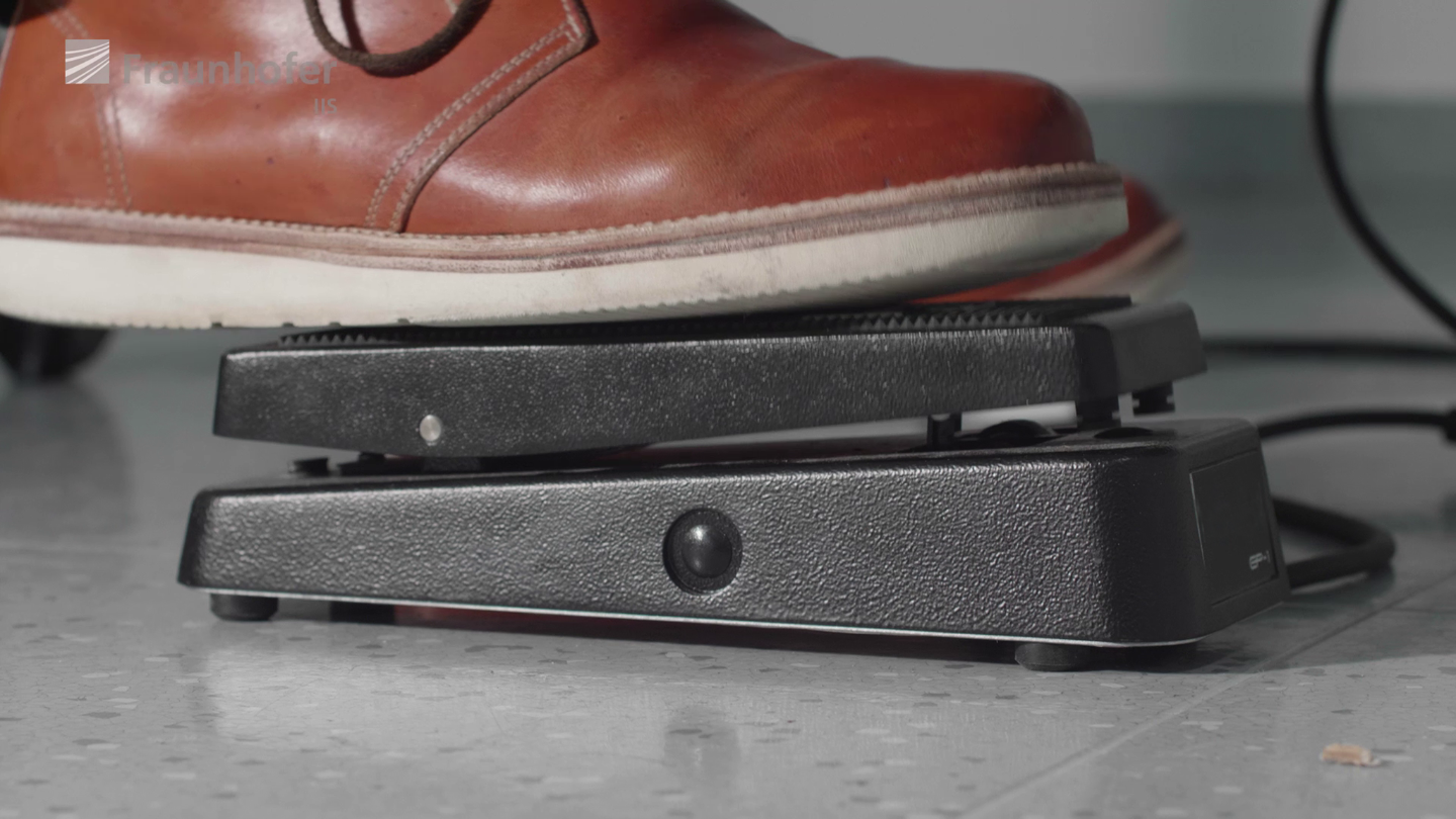
Auto-stereoscopic 3D display, tilted mirror, 2D touchscreen, haptic arm, 3D-printed aspirator, hand motion-capture device, foot pedal, PC
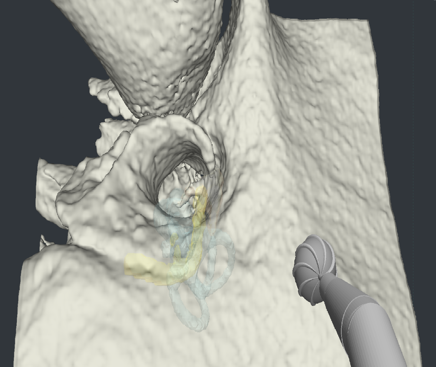
The objective is to drill a hole into the temporal bone behind the ear to access the hearing channel and make space for the cochlear implant (CI) and its electrodes. It is crucial that the surgeon stays clear of the facial nerve and other sensitive structures. The inner and middle ear structures (cochlea, nerves, ossicles, vestibularis) have been segmented from a CT/DVT scan and are highlighted in colors in the virtual 3D model.
Low Running Costs
A virtual simulator has very low running costs compared to one-time usable 3D-printed models, human corpses or animal cadavers
Objective Evaluation
Each session is objectively and quantitatively evaluated. This way
the trainee’s learning curve can be tracked over time
the trainee’s surgical result can be compared against a golden standard created by an experienced expert
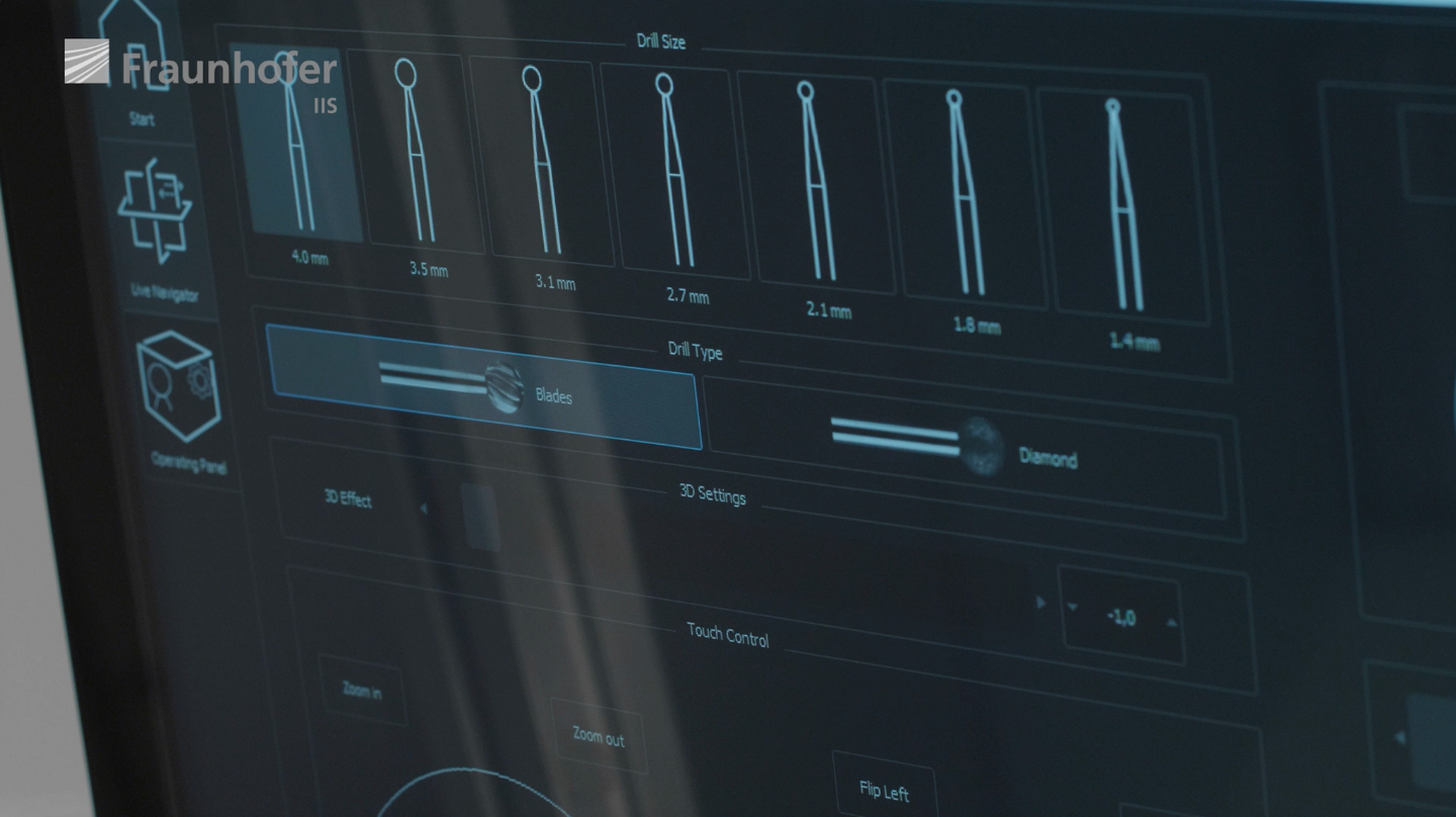
Individualizable Set of Training Cases
Training cases of varying difficulty levels, different pathologies and different patients can be loaded
High Availability
The virtual trainer is always available and does not require lengthy preparations beforehand or cleaning up afterwards. It does not require a lab or surgical theater
The core technology of HandsOn.surgery is the highly realistically modeled interaction between the tool (milling device) and the personalized patient bone model. The underlying physics have been handcrafted by an interdisciplinary team of Fraunhofer IIS scientists and an experienced Ear-Nose-Throat (ENT) specialist. Four other ENT-specialists from multiple hospitals have subsequently evaluated and validated the physical simulation.
HandsOn.surgery is not certified for diagnostics or therapy of diseases. It is not a certified medical product.