Computer Assisted Diagnosis for Mammography

Background

Today mammography is the most effective technique available for the early detection and diagnosis of breast cancer. One of the most difficult tasks in the context of mammogram interpretation is the discrimination of benign and malignant lesions based on their appearance in mammograms. When a lesion is suspected to be malignant, usually a breast biopsy is performed. Several studies have shown that the positive predictive value (PPV) of mammography interpretation usually is not higher than 30 percent. And therefore, only few breast biopsies actually show a malignant pathology. The resulting high percentage of unnecessary breast biopsies on benign lesions causes both considerable mental and physical discomfort for the patients as well as unnecessary costs for the health care system.
Our Approach: MammoCAD

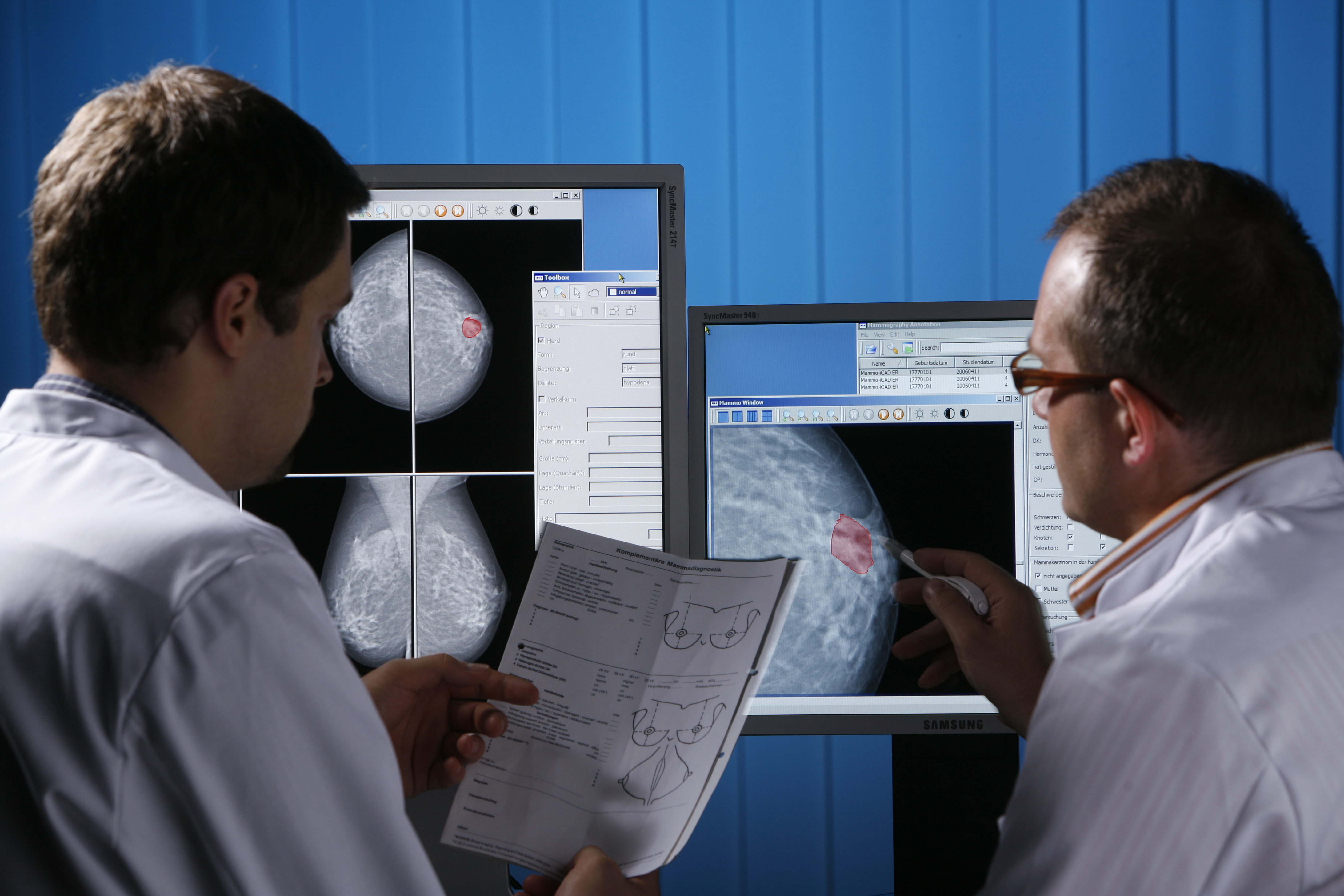
We are working on a system for the computer-assisted discrimination of benign and malignant mammographic masses and clusters of microcalcifications. The ultimate goal of our system is to help the radiologist to improve the reliability of his recommendations for patient management and thus to reduce the high percentage of breast biopsies on benign lesions. Our approach is based on the retrieval of similar lesion information from a validated and annotated database based on the image content and clinical data. Automatic retrieval of similar reference images based on the image content supports the case-based diagnosis during the findings report. This procedure allows for the access of suitable reference cases, including the relevant patient history data during examination.

“MammoCAD“ is not yet CE-certified, as the compliance with requirements of the German Medical Product Law (MPG) and the European Directive 93/42/EEC (MDD) has not been proofed yet. It cannot be obtained before the conformity with the MPG and the MDD has been declared (CE mark). The Fraunhofer IIS is presenting “MammoCAD“ with the objective of gaining partners for further development, production and marketing.

More Information


Your Development Partner for Medical Image Analysis

Talk to us – together we will find a solution for your medical image processing challenge:  -> Please send an email if you are interested

Software based Image Analysis and Enhancement

Our focus are the domains of Microscopy and Endoscopy.