 Fraunhofer Institute for Integrated Circuits IIS
iStix® - A cost-effective scanning solution for microscopy
Erlangen/Düsseldorf, Germany: To generate high resolution panoramas of microscope images, it is often necessary to employ expensive slide scanners. The Fraunhofer Institute for Integrated Circuits IIS has developed iStix®: A scanning solution that combines image processing software and a camera to enable areal scans with your own microscope. iStix® will be presented for the first time at MEDICA in Düsseldorf from November 13 to 16, 2017.
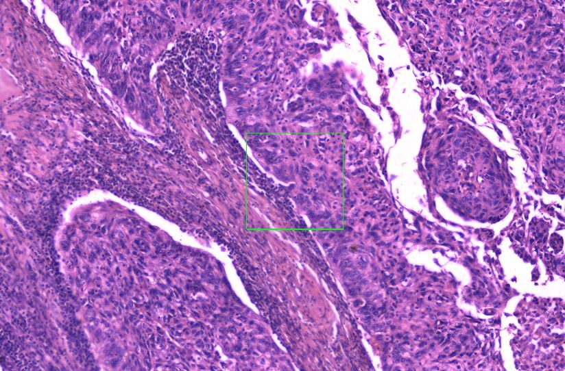
In clinical pathology, medical findings are usually based on microscopy images of tissue sections and other test results. For documentation purposes or a second opinion, a digitized high resolution panoramic image of the examined tissue sample proves extremely valuable. However, often the relevant piece of information consists of individual cells or a morphological region of interest, much smaller than the entire tissue sample. Thus it currently is too time-consuming to scan the entire tissue with a dedicated scanner. With iStix® we offer a scanning solution for pathologists that seamlessly integrates into their microscopes.
Fraunhofer IIS has developed a cost-effective and easy-to-use alternative to digital slide scanners. iStix® is a software solution for generating large-area scans using a manual microscope and a camera. An automated image processing method stitches individual microscopy images together in real time based on the image contents – without the need for a motorized stage. During the stitching process, the images are correctly placed to create a panoramic image, known as a whole-slide dataset. The integrated zoom and annotation functions render iStix® an efficient and simple solution to share data.
High-resolution image data speeds up diagnostics
iStix® generates panoramic images – large-area scans – in real-time, enabling faster diagnostics and an easy way to document findings with a high-resolution overview image. The software is easy to use, extremely versatile and can be integrated into existing applications or pathology and laboratory information systems. iStix® can be easily combined with any microscope that has a camera attached.
Fields of application in clinical diagnostics and materials science
iStix® can contribute to many different applications like telepathology, documentation systems or training of doctors and biologists. Still other fields are conceivable where high-resolution microscopic images play are crucial role, such as material science, quality assurance and material testing, where stress on newly developed materials need to be examined. Within the scope of a development contract, Fraunhofer IIS offers to implement additional customized functions or modifications, as required. Further developments are conceivable in, for instance, the medical field and for image classification and evaluation.
Partners needed for pilot study
The iStix® software is not yet certified as a medical product. For this purpose, Fraunhofer IIS is currently looking for additional partners to evaluate iStix® within the scope of a pilot study. Possibilities for testing the iStix® technology will be presented at MEDICA in Düsseldorf from November 13 to 16, 2017. In Hall 10, Booth G05, visitors can learn more about integrating the technology.