Click-CT: a compact Subμ CT scanner

About Click-CT

© Fraunhofer IIS
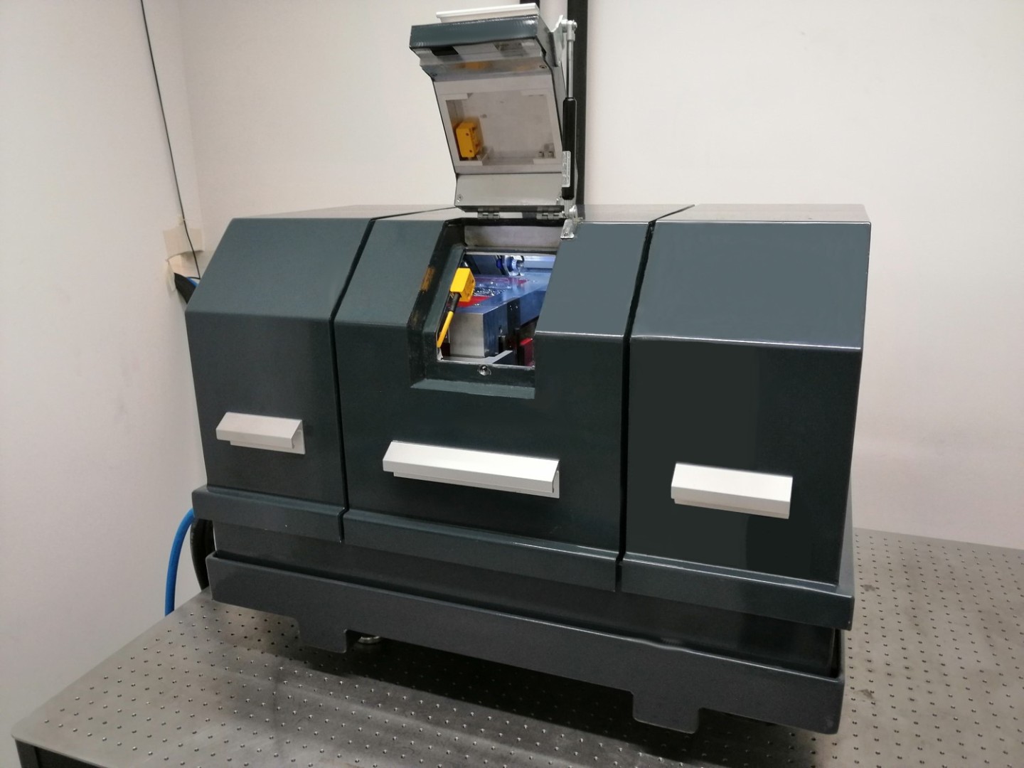
Die Click-CT ist ein kompaktes Sub-Mikrometer (subμ) CT-System.

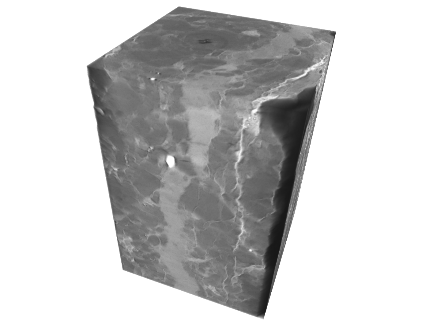
The Click-CT scanner is a compact sub-micrometer (subμ) CT system designed by Fraunhofer EZRT for non-destructive volumetric materials characterization at the highest standard. To achieve high-contrast imaging while acquiring CT data sampled with down to 300 nm voxel-size the user can pick one out of three scintillator screens meeting his requirements. Choosing one of the three magnifications available the fieldof- view can be varied from 0.61 mm to 3.0 mm (up to 5.4 mm using field-of-view extension). Scan quality is comparable to state-of-the-art synchrotron imaging beamlines which employ similar technology as Click-CT.

Microscope X-ray detector

© Fraunhofer IIS
Volumenwiedergabe von Grauwacke-Gestein (J. Tiedemann, TU Berlin), die Probe ist 2 mm dick (4 x Linse, 1,6 μm).

Click-CT draws its resolving power from an indirect microscope detector which has three lenses: 20 x, 10 x and 4 x (see table for the corresponding pixel sampling and field-of-view FoV). The detector features a cooled sCMOS camera with outstanding low noise and high quantum efficiency. Lenses and scintillators can be changed and optimized for any application

Lens Pixel size/μm
Field-of-View/mm (extended)
20 X 0.32 0.61 (1.1)
10 X 0.64 1.2 (2.2)
4 X 1.62 3.0 (5.4)

Automated image processing

© Fraunhofer IIS
Click-CT misst den Drift des Brennflecks in den Objektkoordinaten in situ und korrigiert automatisch die Rückprojektion.
  • Lens focusing and pre-scan geometry calibration are fully automated
  • Signal-to-noise ratio is maximized by automatically adjusting the source-object distance
  • Intuitive parameterization of Paganin-type phase retrieval and and Wiener-deconvolution (both are applied during automated volume reconstruction)

Key applications

© Fraunhofer IIS
  • Short- and long-fiber composites
  • Microstructure of metallic alloys
  • Granular materials and micro foams
  • Detection of microscopic defects (cracks and voids)
  • Determination of tree species from wood samples (dendrology)
  • Detection of ash loading in filters
  • Cathode and anode materials of Li-ion batteries
  • Paper and paper-coatings
  • Biomaterials

Specifications

X-ray source

  • 40 kV – 150 kV peak voltage
  • 4 W – 75 W power

Dimensions

  • L x W x H (in mm) = 900 x 630 x 600

Customized X-ray detector

  • 4 x, 10 x and 20 x motorized lenses
  • 1 μm – 300 μm scintillator screens (typical thicknesses 3 μm, 10 μm and 50 μm)
  • 2048 x 2048 sCMOS camera*

*water-cooling is recommended

Software

  • Step-wise or continuous rotation
  • Automated geometry & detector calibration
  • Focal spot and intensity drift correction in situ
  • Multi-material phase retrieval
  • Field-of-View CT extension (half-scan)