 Fraunhofer Institute for Integrated Circuits IIS
Automated microscope scanner is an all-around talent
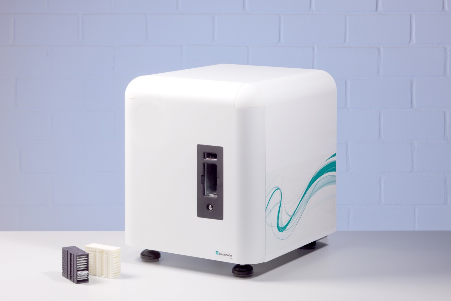
Erlangen/Munich, April 28, 2016 – Analytica, Hall A1, Booth 526: The SCube® is a fully automatic microscopic scanning system developed by the Fraunhofer Institute for Integrated Circuits IIS. Highlights of the system include its suitability for a wide range of analyses, support for both transmitted-light and fluorescence microscopy, attractive pricing and compact size. Because it features a modular design, it can be custom configured to meet individual user needs. Researchers are presenting the scanner at the Analytica trade fair from May 10-13, 2016 in Munich.
In hospitals, large numbers of samples are always being sent to the lab or pathology department for further analysis. These include blood, bone marrow and tissue samples that must all be analyzed at the microscopic level. In small laboratories, this is frequently accomplished using manual methods. While there are some automated microscopy systems currently in use, either these are designed for conducting specialized analyses, which means they cannot be employed universally in all situations, or they are intended for high-volume applications, and their high purchase price makes them all but unaffordable for small and medium-sized laboratories.
An ideal automated solution for smaller laboratories
The SCube® microscopic scanning system bridges this gap. It was developed at Fraunhofer IIS as part of the German Federal Ministry of Education and Research’s LoCAMSSA project, which is now coming to conclusion. The system is unique in that it is an all-rounder offering both high-resolution transmitted light microscopy with immersion oil as well as fluorescence microscopy within a single device. “With our scanning system, users can automatically scan and analyze materials such as blood, bone marrow, and tissue,” says Fraunhofer IIS group manager Dr. Christian Münzenmayer. Furthermore, the system is affordably priced, and at approximately 40 cm in length it is also very compact. Thanks to its modular construction, it can be precisely tailored to the specific needs of individual laboratories. In short, it is an ideal automated solution for small and medium-sized laboratories.
An elegant white casing conceals the sophisticated technology contained within in a user friendly manner. There is an opening through which to insert special cartridges, each containing up to ten slides with samples, into the system. A gripping arm inside the system takes one slide at a time and places it into the beam path between the condenser and the lens. The system now begins the microscopic analysis: it takes single still images of the slide using an integrated camera, places the slide back into the cartridge, and begins the process again with the next slide. Users can specify the analysis to be performed for each sample via software input. Researchers are currently designing a cartridge loading system that can insert up to 20 cartridges into the system automatically, thus allowing for overnight operation.
A variety of modules provides flexibility
Because the system is modular, users and laboratory equipment manufacturers can adapt it to their specific applications. “Aside from the cartridge loading system, one example of this is a module containing several lenses that can be changed as needed,” explains Fraunhofer IIS project manager Dr. Malte Avenhaus. Another module makes it possible to measure fluorescence as well. Here, the sample is stimulated from above using the lens and the fluorescent light emitted by the sample is observed. An additional unique feature is the automatic application of immersion oil. When this oil is applied to the slide containing the sample, the resolution increases – making it possible to discern even more minute details. Currently there are just a few devices that can do this automatically, but such devices are suitable only for highly specialized applications. A web-based platform featuring a viewer allows users to examine the resulting images of individual samples. This starts out as a rough top-down overview and can be zoomed in if a suspicious spot is found in order to see the blood or tissue cells in greater detail.
The prototype of the microscopic scanning system is already complete. Researchers will be presenting this along with other image analysis technologies at the Analytica trade fair from May 10-13, 2016 in Munich (Hall A1, Booth 526). Interested companies can utilize their own image processing methods with the system, or they may also avail themselves of the research and development services provided by Fraunhofer IIS.