 Fraunhofer Institute for Integrated Circuits IIS
We are researching and developing faster and highly functional magnetic resonance techniques that assist physicians in diagnostics and provide patients with shorter examination times.
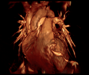
Figure 1: MR reconstruction of a heart.
We are researching and developing faster and highly functional magnetic resonance techniques that assist physicians in diagnostics and provide patients with shorter examination times.
An important acquisition method in magnetic resonance (MR) is parallel imaging and its further development using artificial intelligence (AI). Using multiple transmitting and receiving units simultaneously enables higher acquisition speed. Optimizing parallel image reconstruction with AI makes it possible to obtain high image quality from even less measurement data, which reduces measurement times even more. This opens up new applications that were previously not possible due to not acceptable time requirements.
Conventional image contrast is based on the distribution of water (proton density) and other tissue-specific parameters. The pixel brightness values of an image result from the weighting of these parameters set in the acquisition software. For a comprehensive medical diagnosis of unknown or ambiguous cases often several different contrasts may be needed. Therefore, all possible useful contrasts have to be measured sequentially. Usually, a standard selection of contrasts is used and, in the event of abnormalities, a further examination may have to be performed.
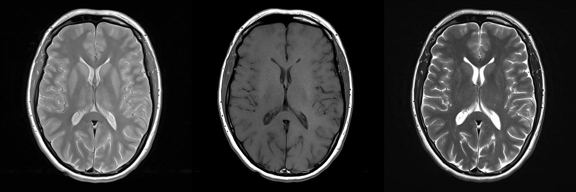
In contrast to conventional examination, quantitative MR takes a holistic approach that requires only one measurement. This measurement does not determine the individual contrasts, but the underlying biophysical parameters of the tissue. The advantage is that, in addition to quantitative parameter values, images with any contrast weighting can be generated by post-processing. Thus, the physician is able to choose the contrast afterwards via software for an optimal diagnosis.