Magnetic resonance imaging in orthodontics

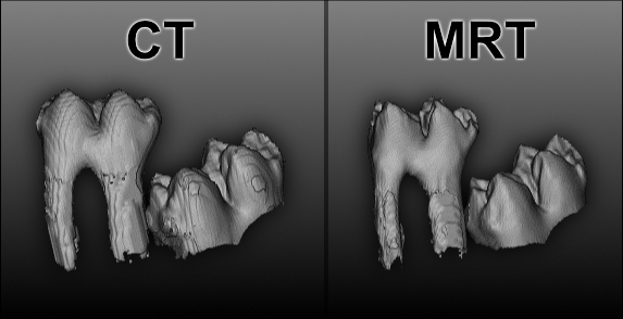
X-ray examinations, i.e. examinations with ionizing radiation, are frequently used for the unequivocal diagnosis of many diseases, including orthodontics. As part of a scientific study conducted by Fraunhofer IIS and Dental Clinic 3 - Orthodontics at Erlangen University Hospital, our researchers were able to show that MRI can be used just as successfully in many areas of orthodontics as the ionizing radiation methods that have been used to date.

In the future, for example, it should be possible to diagnose the position of displaced or impacted teeth without X-rays. The research team received an award for the published results at the annual meeting of the German Orthodontic Society.

Children and adolescents in particular could benefit

MRI procedures can score points with good contrast ratio and the complete absence of ionizing radiation. "The main clientele for orthodontic examinations are children and adolescents. In this group of people, the risk of damage from ionizing radiation is higher than in adults," explains Fraunhofer researcher Dr. Daniel Haddad, referring to a 2007 study.

No significant difference between MRI and other procedures

Figure 1: The study shows that there are no significant differences between magnetic resonance imaging (MRI) without X-rays and computed tomography (CT).

In general, MRI was not significantly different from other methods with respect to most parameters. MRI was even rated better in assessing the location of tooth germs. "We also did not find significant differences in 3D reconstructions of teeth from MRI data compared to conventional methods, except for the surface representation of tooth roots. MRI data acquired with standard parameters appear to be on a par with the X-ray or DVT or CT images used today in many areas" summarizes orthodontist Dr. Andreas Detterbeck of Erlangen University Hospital, adding, "In the area of bone imaging, where X-ray-based methods currently still perform better than MRI, new fast MRI methods offer the chance to close this gap in the future as well."

Research achievement awarded a prize

For the study named "MRI vs.CT for orthodontic applications: comparision of two MRI protocols and three CT (multislice, cone-beam, industrial) technologies" the research team was awarded the annual best prize for the J Orofac Orthop publication 2016 by an independent jury at the annual meeting of the German Society of Orthodontics e.V. on October 11, 2017 in Bonn.