Dr. Haddad, what was the initial idea behind developing this technology? The average person doesn’t really see a difference between X-ray and MRI images.
The main group of patients for orthodontic tests is made up of children and adolescents. Compared to adults, this group is more susceptible to the risk of damage caused by ionizing radiation, as shown by the Brody study from 2007. The advantages of MRI techniques are that they provide good contrast without using any ionizing radiation. An Australian study from 2013 reports that for the group studied, the general risk of developing cancer was 24 percent higher for those people who had a CT scan in the years leading up to the illness than those who had not. So it would be great if in the future we could use alternative examination options for orthodontics that would completely avoid damage caused by radiation.
What has to happen before the technique can be applied for widespread use in orthodontics or even in dentistry generally?
There are various points to consider:
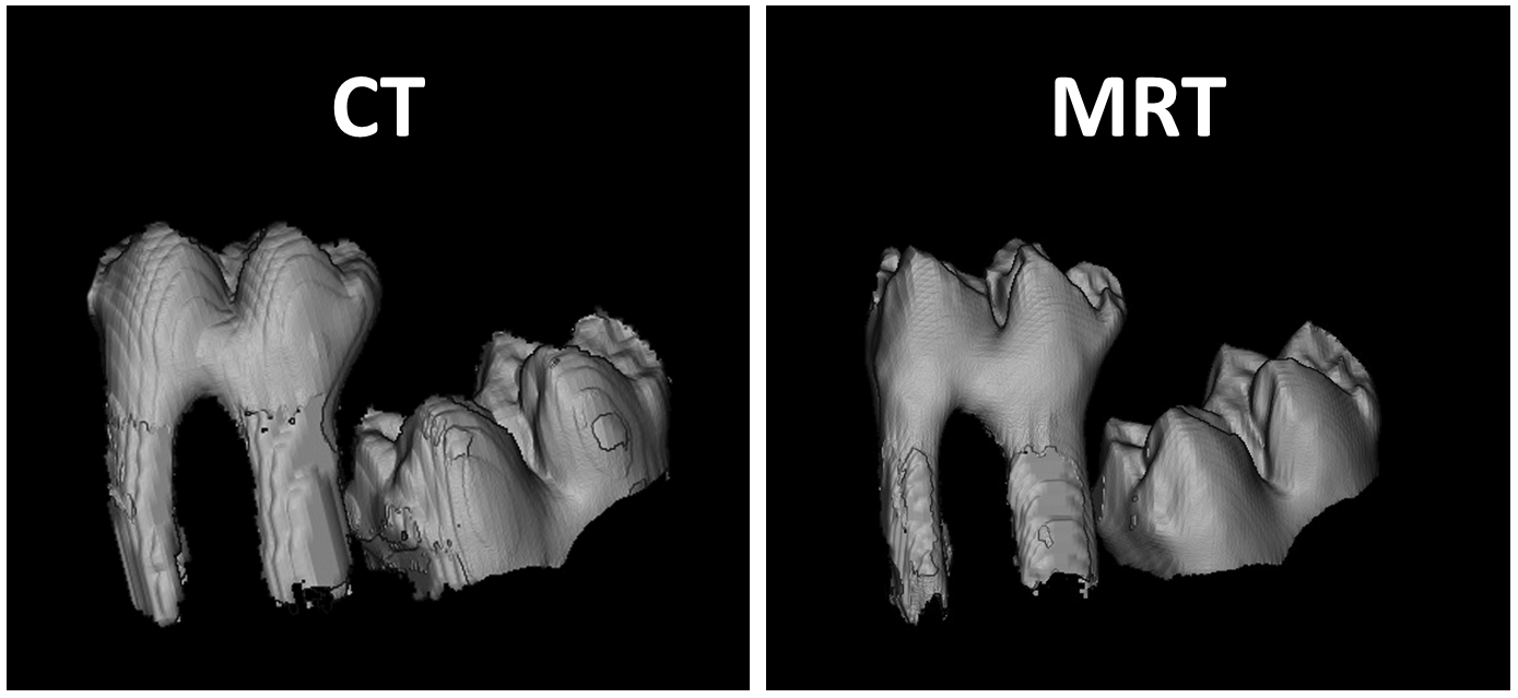
First it’s crucial to significantly raise acceptance for the use of MRIs as a replacement for current techniques that use ionizing radiation. In particular, this entails testing all the sample applications individually to determine the ones in which magnetic resonance imaging is equally good or even better than current methods for diagnosis or treatment. More specifically, it must be shown for each application that the data an MRI offers orthodontists or dentists is at least as conclusive as that currently gained through other methods. It’s also necessary to train physicians properly since MR images can look completely different from the likes or X-rays or CT images delivering the same degree of information.
And second, there is much development work still to be done. This primarily concerns cases in which an MRI can provide as much or even more data than current methods. What we need are MR experiments that are tailored to each point of investigation, but that are also easy to perform, user-friendly and require little training. This may also involve ensuring that when processing the MRI data, image contrast and the way the images look should be similar to those of images generated with current methods.
In addition, MRI machines currently in use would have to be developed and approved for widespread use in orthodontics. Up to now, only typical clinical MRI machines have been used, ones equipped with the necessary detectors, for instance head and jaw coils. This can remain the case in the future and is certainly easy to accomplish in a hospital or university clinic setting, where orthodontics and radiology are on the same site or even same building. Most cases seen at local orthodontic practices would require a referral to a radiologist.
 Fraunhofer Institute for Integrated Circuits IIS