 Fraunhofer Institute for Integrated Circuits IIS
For gastroenterologists, surgeons and any physicians who perform endoscopic procedures, a restricted field of view limits their effectiveness. But if they want to see the full context during a procedure, however, they have to move the endoscope and its connected camera at regular intervals, which means moving it away from the site of intervention. The image the camera captures is visible only temporarily and it is up to the physician to mentally orient themselves regarding the endoscope’s position. And when it comes to documenting the procedure, the only visual material available is individual image frames with a restricted field of view or whole video sequences that are cumbersome to share and archive.
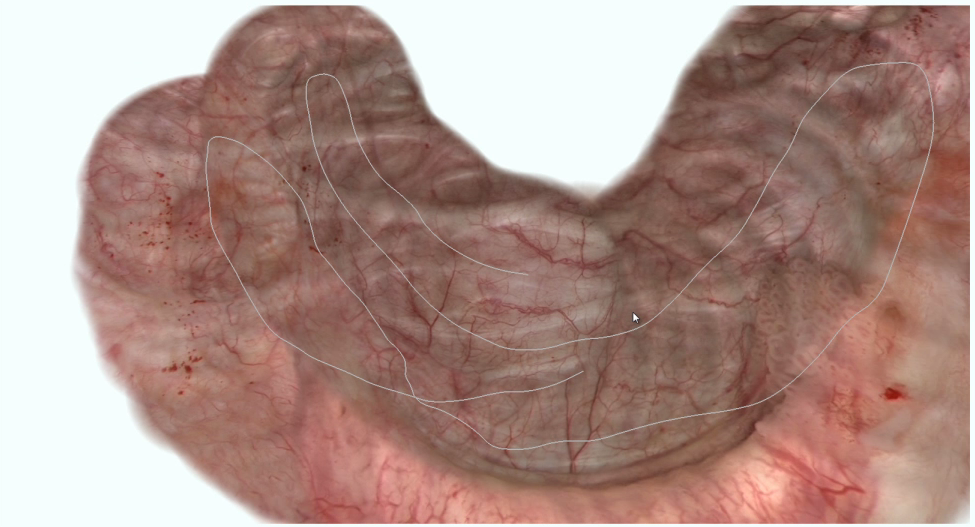
Endorama® - Panorama endoscopy in real time
Your advantages with Endorama®


Rapid orientation thanks to real-time panoramas
- Anatomical context during endoscopic procedures
- Dynamic, expandable field of view achieved through simple movements of the endoscope
- Live image displayed centrally
High-resolution overview for image-based documentation
- Panoramic image can be integrated directly into digital patient files
- Annotations can be inserted, enabling easy information sharing
- Lesions are easier to find again
- Individual images (raw data) are stored in a panoramic image and easily opened by mouse click
- Intuitive controls (pan and zoom) for viewing the panoramic image
Quality control
- Not yet examined areas are displayed as blanks in real time
- Endoscope path is superimposed onto the image
Endorama® Stitching technology
Panoramic image calculation with Endorama® is based on a sequence of image operators, which are applied directly to each image that the connected camera captures. After distortion of the endoscopic image is corrected, image features are used to spatially align the image sequence. In this way, a panoramic image of the scene captured by the endoscope is gradually established with a constant data rate. The most recent image is always projected onto the panoramic image so that the user can constantly see a live image in anatomical context.
Please Note
Endorama® has not yet been certified as a medical product. Fraunhofer IIS is presenting Endorama® with the aim of engaging partners to further develop, manufacture and market it.
Publications and related links
- Bergen, T; Schneider, A; Münzenmayer, C; Knödgen, F; Feussner, H; Wittenberg, T; Winter, C: Echtzeit-Stitching endoskopischer Bilder für eine erweiterte Sicht in chirurgischen Eingriffen. In: Endoskopie Heute, 24(1):60-61, 2011.
- Bergen, T; Ruthotto, S; Rupp, S; Winter, C; Münzenmayer, C: Endoscopic Egomotion Computation. In: Proc’s SPIE Medical Imaging 2010: Computer Aided Diagnosis, Feb. 13-18, 2010, Dawant, BM; Haynor, DR (Eds.), Vol. 7623.
- Endorama®: Development and first clinical experience - European urology supplements Vol.15
- Endorama® panorama endoscopy: 67th congress of the German Association of Urology (german)
- Panoramic imaging of the bladder - endoscopy today (german)
- Software unwraps cystoscope's tunnel view to better image GU tract - medgadget.com
Podcasts
Archive
Here you can find information about finished projects