Contract Development for Medical Image Analysis

Our profile

We are a team of bioinformaticians with many years of experience and a track record in the processing and analysis of medical images, especially in the areas of

  • Microscopy
  • Endoscopy
  • Ophthalmology
  • and in related disciplines.

We have acquired medical expertise through collaboration in interdisciplinary teams (pathology, biology, cytology, hematology, endoscopy, and ophthalmology).
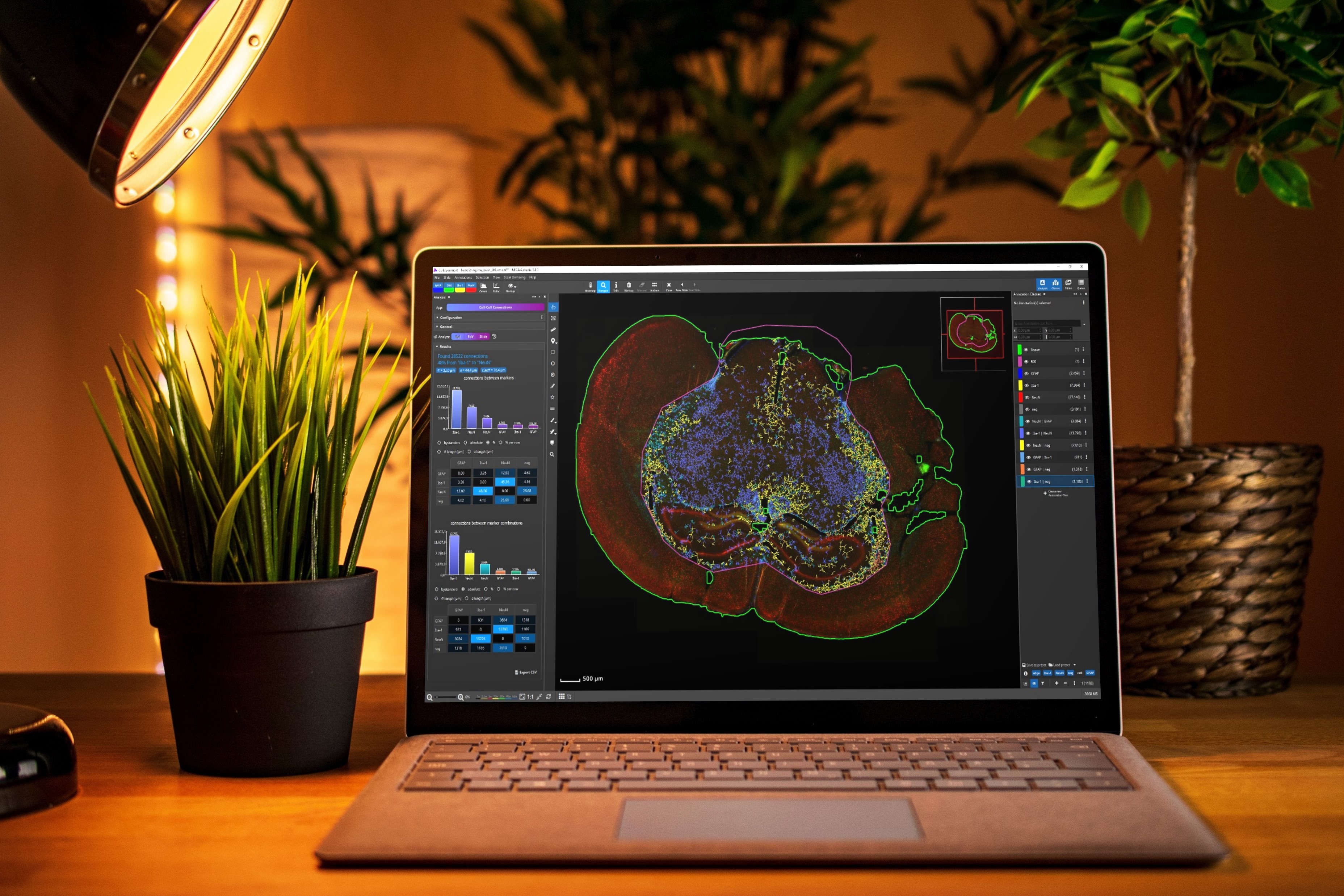
Your advantages with us as an R&D partner

Typically, we are involved in the early stages of medical product development. With a focus on real-time image analysis methods and microscopy and scanning systems, we support you from the first step to complete system conception.


Our application-specific solutions and services include

  • conducting workshops
  • consultation
  • feasibility studies
  • image analysis as a service
  • algorithm and AI development
  • software engineering of applications or libraries
  • customization and re-branding of our existing software


Our expertise includes

  • (Classical) computer vision and AI (CNNs, transformers)
  • Development of new algorithms
  • Training of new AI models
  • Code optimization for speed (GPGPU, Multi-Threading, cloud)


Reference projects for the international industry

  • Software development for a pathology scanner distributor (real-time JPEG 2000 image compression on GPU, whole-slide image creation, viewer, annotation system)
  • Research and development of AI and software for primary image analysis for a device manufacturer in the field of spatial biology
  • Development of image analysis software for a fluorescence microscope and scanner vendor
  • Research and development of real-time 3D colon reconstruction for monoscopic colonoscopies for a clinical documentation system provider
  • Research and development of real-time polyp detection and classification for monoscopic colonoscopes for clinical documentation system vendors.
  • Development of real-time video optimization for fiber optic endoscopes for an endoscope manufacturer, executable on legacy hardware
  • Concept and architecture design of a novel AI-assisted digital endoscopy tower for an endoscope manufacturer
    Statistical analysis and evaluation for the spread of nasal spray in the oral and nasal cavity for a CRO
  • Development of an algorithm to extract high-quality fundus images using a handheld digital ophthalmoscope video (iPhone) for an ophthalmology provider
  • Feasibility study for an AI-based identity verification between iris images acquired with different devices at different spectra and light settings for an ophthalmology vendor
  • Autofocus research and development and feasibility study for real-time 3D stitching for a digital stereo colposcope vendor


Our infrastructure

Digital Pathology Lab

  • Microscopy: Zeiss Axio Scan.Z1, Motic EasyScan, PreciPoint M8 scanner, Fraunhofer SCube, Zeiss AxioImager FL microscope (additional scanners available through our clinical partners).
  • Annotating and Calculating: professional image analysis solutions (MIKAIA , HALO from Indica Labs), WACOM tablet for slide annotation, SlideMatch for registration of consecutive or "re-stained" sections.

Endoscopy Lab

  • Several mono- and stereo-, rigid and flexible endoscopes (Wolf, Storz, Schölly, Olympus), various light sources, robotic arm
  • Various phantoms
  • 3D printer

Deep Learning Cluster: 34 nodes, 72x NVidia P100 GPU, 16x NVidia P40 GPU, 2.8 TB RAM

Extensive partner network from clinics, universities and industry