 Fraunhofer Institute for Integrated Circuits IIS
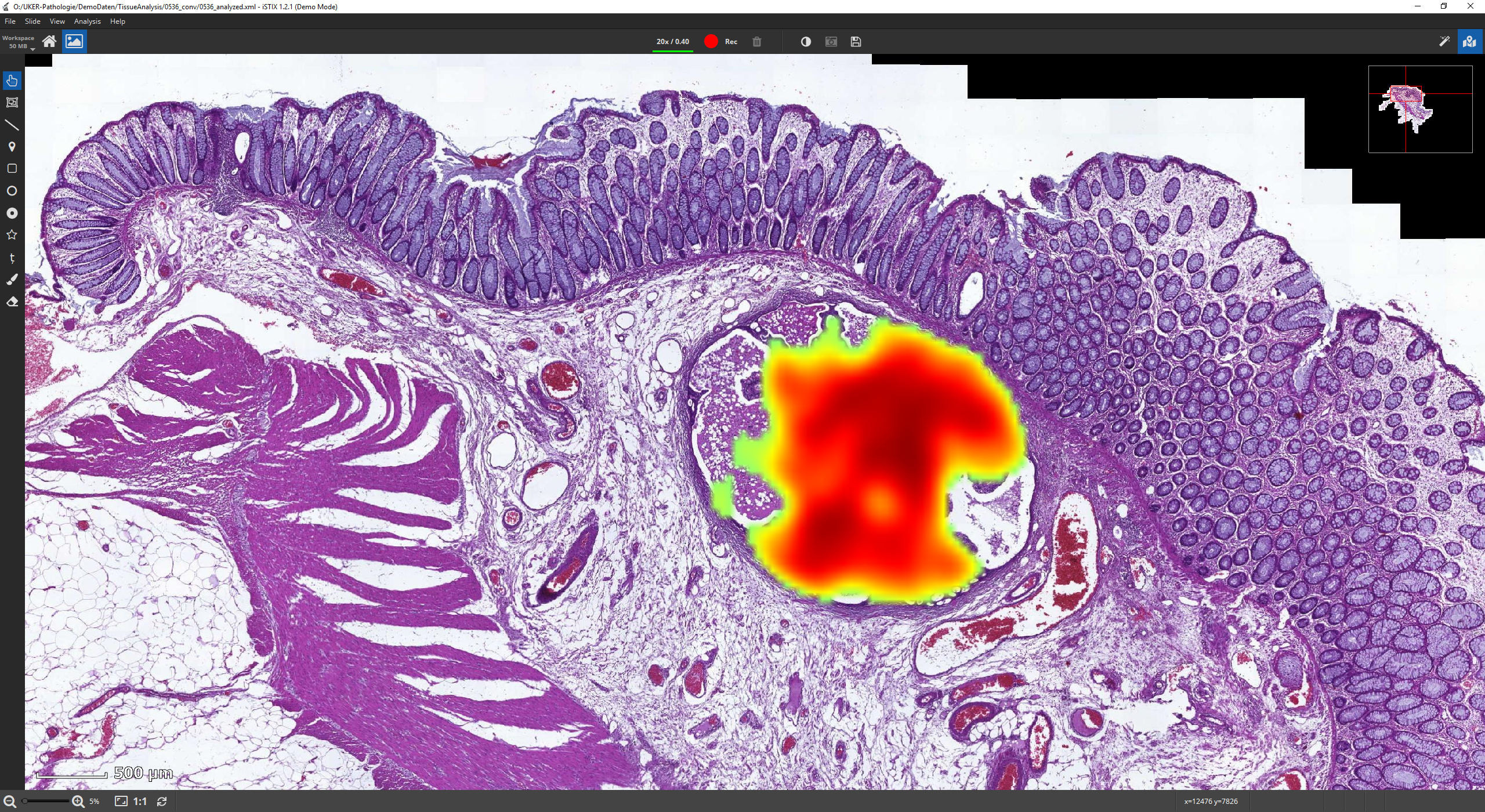
AI-based analysis methods combined with high-resolution microscopy technologies have opened up new possibilities for the analysis of microscopic images.
Our team specializes in developing software solutions and algorithms for processing, as well as qualitative and quantitative evaluation of microscopic images.
We also provide customized services and contract development tailored to specific applications and customer needs.
If you are interested in collaborating on a development project, please don't hesitate to contact us!